4D Mammography Pilot Study Complete, Analysis Underway
Calidar Completes Enrollment in First-in-Human Study of 4D Mammography System with 61 Patients Imaged.
Research Triangle Park, NC – April 28, 2026 – Calidar, Inc., an innovative start-up in precision diagnostic imaging, today announced the completion of enrollment in its first-in-human clinical study of the 4D Mammography system. The study, conducted in collaboration with Baptist Health Hardin in Elizabethtown, KY, enrolled 61 patients and represents a significant milestone in the development of a next-generation imaging platform designed to improve breast cancer diagnostics.
The 4D Mammography system leverages X-ray diffraction to measure molecular-level signals within breast tissue — a capability not available in conventional mammography. With enrollment now complete, Calidar and its clinical partners will proceed with data analysis to evaluate the system's ability to distinguish between healthy tissue and breast cancer. "First-in-human is the gate that separates promise from evidence," said Dr. Carpenter, CTO of Calidar. "We cleared it — and the early performance data is directionally aligned with our clinical thesis, with early signals pointing to up to 4x improvement in precision over 3D mammography. Full analysis is ongoing, but we now have human evidence that suggests 4D Mammography works. Everything that follows is about scale."
The first-in-human study was initiated in August 2025, when the team successfully imaged the first patient with the 4D Mammography system. The completion of enrollment is the next major step in what Calidar believes could redefine how breast cancer is detected at the point of imaging.
Breast cancer diagnostics remain a significant clinical challenge. In the United States, approximately 1.8 million breast biopsies are performed each year, with 70-80% ultimately diagnosed as benign. These unnecessary procedures contribute to over $7.4 billion in annual healthcare costs and place a growing burden on radiologists and pathologists. At the same time, inconclusive imaging results contribute to delayed diagnoses for an estimated 60,000 breast cancer patients annually. By measuring the structure and composition of tissue—not just its shape or density—the 4D Mammography system aims to provide a new dimension of diagnostic clarity.
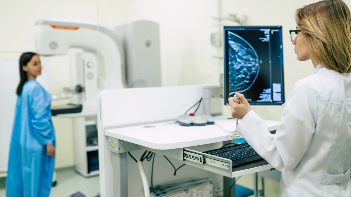
Baptist Health Hardin served as the clinical research site for this study. The study enrolled patients who presented with findings requiring further evaluation. Once enrolled, participants received a 4D Mammogram to test the system’s ability to differentiate between healthy and cancerous breast tissue and benchmarks performance against existing mammography devices. Participation in the study was conducted under investigational use; results were not used to inform individual patient clinical care. Data analysis is ongoing, and performance results have not yet been disclosed. It is anticipated that a peer reviewed paper on the study will be published in the second half of 2026.
To support the final phase of developing 4D Mammography, Calidar is initiating a Series A raise that will fund a multi-site pivotal study, nationally. Following the conclusion of this pivotal study, Calidar anticipates submitting these results in pursuit of FDA approval for 4D Mammography.
The 4D Mammography system is investigational and has not been cleared or approved by the U.S. Food and Drug Administration. It is not available for commercial sale and is limited to investigational use in the United States under FDA’s abbreviated IDE requirements.
About Calidar
Calidar, Inc. is a medtech company developing the world's first clinical application of volumetric X-ray diffraction imaging for breast cancer diagnostics. Its 4D Mammography system reads the molecular fingerprint of breast tissue — delivering high-precision diagnostic information that today's 3D imaging cannot provide. Calidar's technology is designed to reduce unnecessary biopsies, improve diagnostic accuracy, and enable earlier detection at the point of imaging. The company is headquartered in Research Triangle Park, NC.